Facet joints connect the vertebrae the bones of the spine. The following descriptions only summarize the basic anatomic information that every thoracic surgeon and chest physician must know.
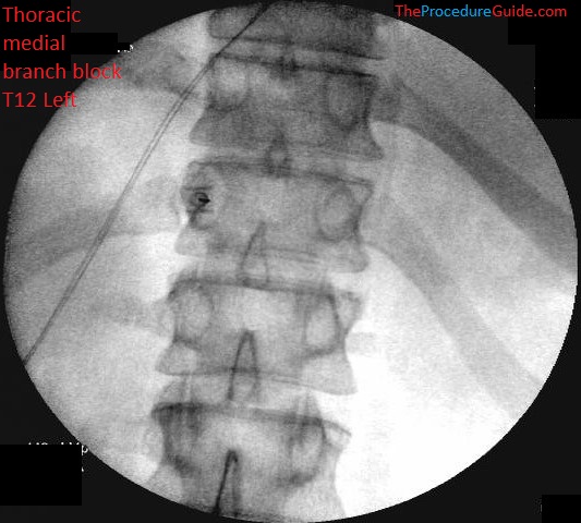
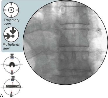
Fluoroscopic Guided Thoracic Medial Branch Block Technique And Overview The Procedure Guide
Up to 10 cash back The medial branches of the thoracic dorsal rami were found to assume a reasonably constant course.

. The origin of the nerve directly from the middle trunk of the brachial plexus has also been reported. Anterior intercostal branches diverge laterally from the internal thoracic artery to run into the first six intercostal spaces. The cervical facet joints guide motion in the neck and the thoracic facet joints guide motion in the mid-back.
Spinalis is the most medial of the three erector spinae muscles. Thoracic Medial Branch Blocks and Cooled RF Coolief The role of thoracic facet joints in chronic back pain has received very little attention as compared to lumbar and cervical facet joints. They supply the intercostal pectoral muscles and the adjacent skin anastomosing with their posterior counterparts near the posterior trunk.
To establish the anatomical basis for thoracic medial branch neurotomy an anatomical study was undertaken. What are thoracic facet joints. The medial branches ramus medialis.
Ultrasonographic Anatomy of the Brachial Plexus and Major Nerves of the Canine Thoracic Limb. The thor-acic aorta is divided into ascending transverse and descending portions Fig. These injections are performed with the use of a posterior approach.
Upon leaving the intertransverse space they typically crossed the superolateral corners of the transverse processes and then passed medially and inferiorly across the posterior surfaces of the transverse processes before ramifying into the multifidus muscles. Thoracic spinal pain can be as chronic and disabling as neck and low back pain even though it is less common. Prior to the steroid injection you will be lying on your stomach.
Feb 22 2022 the lateral. Internal branch of the posterior divisions of the upper six thoracic nerves run between the Semispinalis dorsi and Multifidus which they supply. The medial branches of the posterior divisions of the upper six thoracic nerves run between the semispinalis dorsi and multifidus which they supply.
First branch off of the dorsal side of the spinal nerve. A thoracic medial branch block is an outpatient procedure for diagnosing and treating upper and middle back pain. SURGICAL ANATOMY A detailed knowledge of surgical anatomy is of major importance in the practice of thoracic surgery.
They then pierce the Rhomboidei and Trapezius and reach the skin by the sides of the spinous processes. The medial branches of the thoracic dorsal rami were found to assume a reasonably constant course. The medial branches of the lower six are distributed chiefly to the.
The medial branches of the thoracic dorsal rami were found to assume a reasonably constant course. There are two main patterns of branching of this. A local anesthetic.
A thoracic medial branch block is a diagnostic treatment intended to determine whether a particular thoracic facet joint is the source of your pain. They then pierce the rhomboidei and trapezius and reach the skin by the sides of the spinous processes. For example the T5 and T6 medial branch nerves must be denervated to treat pain that originates from the T6-T7 zygapophysial joint.
General sense touch pressure pain heat cold etc to the skin of the back. Ultrasonography of the brachial plexus including ally the ventral branch of. The posterior ramus of the thoracic nerve passed through the narrow space between the bony structures and adjacent fibrous tissue.
Perforating branches stem from the medial aspects of the internal thoracic artery. They have a short. They help guide your spine when you move.
Detailed anatomy of the posterior ramus and mediallateral branches and their fine branches in the entire thoracic region was investigated by both macroscopic and stereomicroscopic dissections. The medial pectoral nerve may arise directly from the anterior division of the inferior trunk of the brachial plexus. Thoracic medial branch nerve injections are indicated for the diagnosis of axial thoracic ie mid back pain that typically originates from zygapophysial ie facet joint sprains contusions or osteoarthritis.
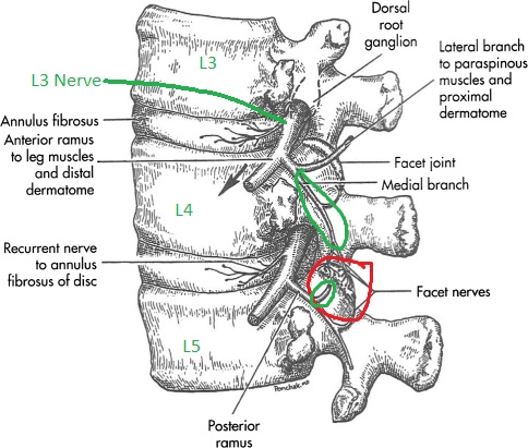
Sympathetic innervation to the skin. It is sent to the first branch which is. The medial branch then runs around the lateral border of the superior articular process and enters the fibro-osseous canal.
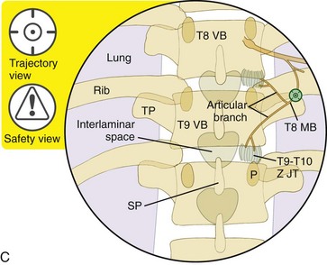
The needle tip is placed at the superior lateral edge of the transverse process where each. At that time however there were no antece- elacque 4 offers a more detailed description of the dent studies of the anatomy of the target nerves which thoracic dorsal rami but again explicit details as to were the articular branches arising from medial radiological landmarks for the medial branch or its branches of the lumbar dorsal rami. What happens during an RFA.
Each vertebral segment has two facet joints one on each side. And median nerves are identified on the medial aspect of mid-humerus and followed proximally to the axillary region and distally to the elbow. Thoracic medial branch anatomy Written By marcou Wednesday March 9 2022 Add Comment Edit.
The area of the spine between your neck and low back is called the thoracic region. A mixed nerve containing both motor and sensory fibers. The medial pectoral nerve arises from the medial cord of the brachial plexus with C8 and T1 nerve roots.
Chest Wall The bony thorax includes the sternum 12 pairs of ribs and costal cartilages and 12. Cervical or Thoracic Medial Branch Block Facet Nerve Injections The facets are the small bony joints that connect one spine vertebra to another at the back of the spinal canal. First branch off of the ventral side of the spinal nerve.
As you progress up the thoracic spine the medial branch nerves sit more lateral on the transverse. This chapter will focus on the radiofrequency technique for neurolysis of the thoracic medial branch nerve. In vertebrates thoracic vertebrae compose the middle segment of the vertebral column between the cervical vertebrae and the lumbar vertebrae.
The medial branches of the lower six are distributed chiefly to the multifidus and longissimus occasionally. The site of the. The thoracic diaphragm or simply the diaphragm ancient greek.
Linton et al estimated the prevalence of all spinal pain in the general. They increase in size going towards the lumbar vertebrae with the lower ones being a lot larger than the upper. Using an X40 dissecting microscope a total of 84 medial branches from 7 sides of 4 embalmed human adult cadavers were studied.
The medial branch runs in the groove formed by the lower transverse process and the superior articular process and then descends caudally and posteriorly accompanying the vessels arising from the lumbar artery and vein. See cervical thoracic and lumbosacral medial branch nerves For example if the allowance in the claim is for the c5c6 joint then the c5 and c6 medial nerve branches are the appropriate targets while an l4l5 joint would involve the l3 and l4 medial nerve branches. Upon leaving the intertransverse space they typically crossed the superolateral corners of the transverse processes and then passed medially and inferiorly across the posterior surfaces of the transverse processes before ramifying into the multifidus muscles.
Two medical branch nerves must be denervated to render a thoracic zygapophysial joint painless. To the deep back mm. To a thoracic medial branch nerve the nerve can no longer transmit pain from an injured facet joint.
The thoracic spine is fairly superficial in many thin patients so you can often get to it with a basic. Duration Less than 15 minutes How is it performed. Facet Joint Cartilage Medial Branch Nerves Facet Joint Capsule Degenerated Facet Joint Normal Anatomy of the Thoracic Spine Degenerated Thoracic Facet Joints Thoracic Facet Joint Pain Patterns.
Fluoroscopic Guided Thoracic Medial Branch Block Technique And Overview The Procedure Guide.

Illustration Of Variation Of Position Of Medial Branch In Thoracic Download Scientific Diagram
Thoracic Zygapophysial Joint Nerve Medial Branch Injection Posterior Approach Radiology Key
Illustration Of Medial Branch Mb And Lateral Branch Lb Thoracic Download Scientific Diagram

Thoracic Facet Injections
Thoracic Zygapophysial Joint Nerve Medial Branch Injection Posterior Approach Radiology Key

Illustration Of Variation Of Position Of Medial Branch In Thoracic Download Scientific Diagram
Fluoroscopic Guided Thoracic Medial Branch Block Technique And Overview The Procedure Guide

Thoracic Facet Injections
0 comments
Post a Comment